 ©
©


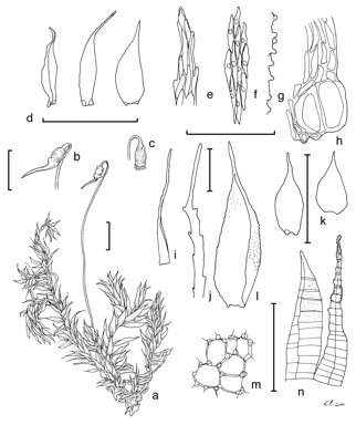
Caption: A, gametophyte bearing sporophyte. B, capsule with operculum. C, capsule showing peristome. D, branch leaves. E, leaf apical cells. F, mid-laminal cells. G, side view of papillae. H, alar cells. I, perichaetial leaf. J, enlarged apex of perichaetial leaf. K, more ovate perichaetial leaves from another specimen. L, ?K? enlarged to show papillose surfaces. M, exothecial cells. N, peristome: folded endostome segment (left) and narrower exostome tooth (A?H, M, N, W.B.Schofield 90362 NSW; I?L, H.Streimann 57010 CANB). Scales: 1 mm for habit, leaves, capsule; 100 mm for cellular drawings including peristome.
Reproduced from H.P.Ramsay, W.B.Schofield & B.C.Tan, The Journal of the Hattori Botanical Laboratory 95: 44, fig. 18 (2004).Illustrators: L. Eklan & H.P. Ramsay
Australian Mosses Online